What is pigmentation?
All skin colour (pigment) is a result of melanin, two different substances produced by specialised cells in the skin called melanocytes. Melanin functions to protect skin by absorbing and dissipating UV radiation, thereby preventing free radical formation and skin damage. Melanin is essentially our natural sunscreen (fun fact: it has an SPF of 3.4, meaning it absorbs about 50-75% of UV light hitting the skin).
Melanocytes are found in the basal (deepest) layer of the epidermis. They synthesise and package melanin inside the cell using an enzyme called tyrosinase and transfer the melanin-containing package (called a melanosome) to between 30 and 40 neighbouring skin cells (keratinocytes) using finger-like extensions called dendrites. These is how melanin is distributed throughout all layers of the epidermis.
Melanin produces all pigment in normally functioning skin and hair, but mottled brown patches of pigmentation (called hyperpigmentation) are aesthetically displeasing, and usually one of three types:
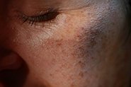
Photo-ageing related pigmentation: caused by exposure to UV light.
Melasma: a complex pigmentary disorder thought to involve a hormonal influence, but triggered by UV exposure.
Postinflammatory hyperpigmentation (PIH): caused by trauma (injury or inflammation) to the skin, particularly in people with darker skin tones.
Pigmentation makes us look older!
Cosmetically, we don’t like looking at our own patchy skin tone in the mirror, but there is evidence that points to hyperpigmented skin being assessed as ‘older’ than an even skin tone.
Multiple experiments have concluded that 3D simulated faces (both male and female), standardised to vary only in skin colour distribution (i.e. even skin tone vs pigmentation), signalled youth, health and attractiveness when they have an even skin tone versus mottled appearance due to age and UV induced photodamage. This was completely independent of wrinkles, skin laxity and loss of facial volume, all things that are typically considered ‘ageing’.
Photoaging and UV induced pigmentation
When melanocytes are exposed to UV, they upregulate the enzymes involved in melanin synthesis and transfer more melanosomes to more skin cells. UV also damages melanocyte DNA, resulting in increased melanin synthesis. The skin cells themselves also respond to UV radiation by taking up more melanosomes, and producing inflammatory chemicals.
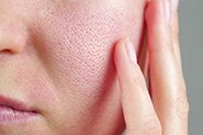
All of this results in pigmentary changes in the skin. Aged skin that has been protected from the sun is pale and wrinkled, but generally with regular, not irregular, pigmentation – think about the skin on your torso that doesn’t see as much sun vs the skin on your arms, chest, face and hands (check out the image if that’s not frightening enough for you!).
Note: Photoaging from chronic UV exposure is really a minor cosmetic concern, although it can significantly affect our self-confidence. The much larger concern with UV damage is DNA damage caused by UV radiation – if this exceeds our cells’ own DNA repair mechanisms, it results in mutations and immune system changes that are the basis of skin cancer and melanoma development.
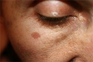
Melasma
Melasma is patchy brown hyperpigmentation that occurs on sun-exposed areas of the face. It can also appear on the neck and arms, usually in post-menopausal women. You may have heard it called ‘the mask of pregnancy’ because it often appears in pregnancy, however oral contraceptives and hormone replacement therapy can also be involved. Sun exposure is a major triggering and aggravating factor.
Melanocytes contain oestrogen receptors, so it is thought that the melanocytes in people with melasma are more sensitive to the stimulatory effects of oestrogens. This results in larger melanocytes that produce and transfer more melanosomes to keratinocytes, resulting in hyperpigmented patches.
Unlike UV-induced hyperpigmentation which appears as random patches, melasma appears in one of 3 patterns:
Centrofacial (most common): a symmetrical pattern on forehead, cheeks, upper lip, nose and chin
Malar: the cheeks and nose
Mandibular: the jawline
Melasma is a frustrating and emotionally distressing disorder as it is chronic and often relapses, especially with continued sun and hormone exposure.
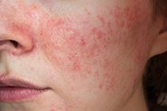
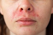
Post-inflammatory Hyperpigmentation
PIH is a reaction to injury or inflammation that causes the skin to produce increased pigment. It is more common in darker-skinned individuals. The most common cause of PIH is acne, but it also can result from psoriasis, a burn, or any skin injury.
PIH results from a local overproduction of melanin and appears as dark spots/patches in the same area as the healed injury. These marks can take several months to years to resolve, even with treatment. And they may worsen with exposure to UV. This is why PIH is THE major reason you shouldn’t pick at your face, and wear sunscreen if you suffer from breakout-prone skin!
IF YOU WOULD LIKE TO DISCUSS PIGMENTATION TREATMENT OPTIONS, CLICK HERE TO BOOK A COMPLIMENTARY CONSULT WITH OUR EXPERIENCED MEDICAL TEAM.
Want to get started? Click here >
REFERENCES
1. Bolognia JL and Orlow SJ, 2018, Melanocyte Biology, New York: Elsevier Ltd, Clinical Key.
2. Cario M, 2019, How hormones may modulate human skin pigmentation in melasma: an in vitro perspective. Exp Dermatol, Mar 18 [epub ahead of print].
3. Chaowattanapanit S, Silpa-Archa N, et al. 2017, Postinflammatory hyperpigmentation: a comprehensive overview: treatment options and prevention. J Am Acad Dermatol, 77(4): 607-621.
4. Chopra K, Calva D, et al. 2015, A Comprehensive Examination of Topographic Thickness of Skin in the Human Face. Aes Surg J, 35(8):1007-1013.
5. Dessinioti C, Lotti TM, et al. 2015. Melasma. In: Katsambas AD, Lotti TM, Dessinioti C, D’Erme AM (eds) European Handbook of Dermatological Treatments. Springer, Berlin, Heidelberg
6. Ebanks JP, Wickett R, et al. 2009, Mechanisms regulating skin pigmentation: the rise and fall of complexion coloration. Int J Mol Sci, 10(9): 4066-4087.
7. Fink B, Grammar K, et al. 2006, Visible skin color distribution plays a role in the percpeption of age, attractiveness, and health in female faces. Evol Hum Behav, 27(6):433-442.
8. Fink B, Matts PJ, et al. 2018, The effect of skin surface topography and skin colouration cues on perception of male facial age, health and attractiveness. Int J Cosmet Sci, 40(2):193-198.
9. Gillbro JM & and Olsson MJ, 2011, The melanogenesis and mechanisms of skin-lightening agents – existing and new approaches. Int J Cosmet Sci, 33:210–221.
10. Gordon J & Brieva J, 2012. Unilateral Dermatoheliosis. New Engl J Med, 366(16):e25-26.
11. Kaufman BP, Aman T, et al. 2018, Postinflammatory hyperpigmentation: epidemiology, clinical presentation, pathogenesis and treatment. Am J Clin Dermatol, 19(4):489-503.
12. Sarkar R, Arora P, et al. 2013, Cosmeceuticals for hyperpigmentation: what is available? J Cutan Aesthet Surg, 6(1): 4-11.
13. Sonthalia S, Jha AK, et al. 2017. Dermoscopy of melasma, Indian Dermatol Online J. 8(6):525–526.
14. Taylor SC, 2005, Photoaging and Pigmentary Changes of the Skin. Berlin: Springer, Springer Link.
15. Trivedi MK, Yang FC, et al. 2017, A review of laser and light therapy in melasma. Int J Womens Dermatol, 3(1):11–20.
16. Zubair R, Lyons AB, et al. 2018. What’s new in pigmentary disorders. Dermatol Clin, 37(2):175-181.